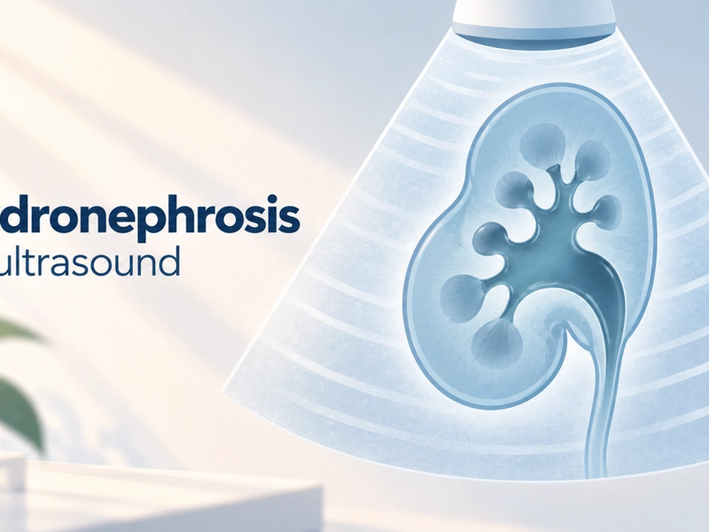
An anechoic cyst on your ultrasound simply means a small pocket of clear fluid that shows up completely black on the scan, and that blackness is usually good news. Let us break the word down. A cyst is a sac filled with fluid, a bit like a tiny water balloon. Anechoic is made of two parts: an, meaning without, and echoic, meaning echoes. So anechoic means without echoes. Ultrasound works by bouncing sound waves off tissues, and clear fluid does not bounce them back, so it appears as an even black area on the screen. That is exactly what a simple, harmless cyst looks like. In other words, the word that sounds most technical on your report is often the one telling you the cyst is the reassuring, clear-fluid kind. Anechoic cysts turn up in the ovary, kidney, breast, thyroid, liver and elsewhere. On your report, anechoic is a description of what the scan showed, not a diagnosis.
If a scan report just landed on your WhatsApp and the word anechoic is making you anxious, slow down, because it is usually one of the more reassuring words on the page. This guide explains what it means, why it is a good sign, and what to ask your doctor. And if the rest of the report still reads like another language, FlexReport can help. Upload it on app.flexreport.ai and get a simplified, plain-language version of the whole thing in minutes.
What does anechoic cyst mean?
An anechoic cyst is a fluid-filled sac that appears completely black on ultrasound because the clear fluid inside it reflects no sound waves. Ultrasound builds a picture from echoes: solid tissues bounce sound back and show up as shades of grey, while clear fluid lets sound pass straight through and returns nothing, which the screen shows as black. Radiologists, the specialist doctors who read scans, use the word anechoic for exactly this even, echo-free blackness. It is the classic appearance of a simple cyst, the most common and most harmless kind. Because clear fluid is so easy for ultrasound to recognise, an anechoic description is one of the ways radiologists reassure themselves that a cyst is the ordinary, benign type rather than something that needs a closer look (Radiological Society of North America).
Why anechoic is usually a reassuring word
Anechoic is usually reassuring because it points to clear fluid, and clear fluid is the signature of a simple, benign cyst rather than a solid growth. When radiologists worry about a lump, it is generally because it looks solid, mixed, or has bright specks and dividing walls inside, none of which fit a smooth black anechoic area. So while the word looks intimidating, it is often doing the opposite of raising alarm. That said, anechoic describes only what the fluid looks like, not the whole story. The size of the cyst, where it sits, and whether it causes symptoms still matter, and those are for your doctor to weigh. The table below shows how an anechoic simple cyst compares with the kind of appearance that gets a closer look.
| Feature | Anechoic (simple) | Complex (not anechoic) |
|---|---|---|
| How it looks | Even, completely black | Uneven, grey specks or bright spots |
| Inside | Clear fluid only | Fluid plus solid bits, walls or debris |
| Usual meaning | Almost always benign | Usually still benign, but reviewed more closely |
| Typical next step | Often no action, or one repeat scan | A follow-up scan, sometimes more tests |
Anechoic cysts in different parts of the body
The word anechoic means the same thing wherever it appears, but what a simple cyst tends to mean can vary a little by organ, so it helps to see it in context. In the ovary, anechoic cysts are extremely common and often just part of the monthly cycle. In the kidney, a simple anechoic cyst is one of the most frequent findings as we age and is usually harmless. In the breast, an anechoic cyst is typically a benign fluid pocket. In the thyroid and liver, anechoic cysts are common and usually need no action. Across all of them, anechoic is the reassuring part of the description. The table gives a quick sense of each, but your doctor always reads the finding in your specific context.
| Where it is found | What an anechoic cyst usually means there |
|---|---|
| Ovary | Very common, often a normal part of the cycle. Read alongside size and symptoms. |
| Kidney | A common simple cyst, usually harmless, especially with age. |
| Breast | Typically a benign fluid pocket. Your doctor confirms with the full picture. |
| Thyroid | Common and usually harmless. Watched if larger. |
| Liver | A common simple cyst, usually needs no treatment. |
The most technical-sounding word on your report is often the most reassuring. Anechoic just means clear fluid.
Is an anechoic cyst serious? Is it cancer?
An anechoic cyst is almost never cancer and is usually not serious. The whole point of the word is that it describes clear fluid, and clear-fluid cysts are the benign, ordinary kind. Cancers and other growths that warrant attention tend to look solid or mixed, not smoothly black. That is why an anechoic description generally lowers concern rather than raising it. A small number of cysts do need follow-up, usually because of their size, a change over time, or symptoms they cause, but that is about the whole picture, not the word anechoic. What matters is your age, where the cyst sits, its size and whether it troubles you, and putting that together is your doctor's job, not a search engine's. A report line is one puzzle piece. Your doctor holds the whole puzzle.
What happens next after this finding?
For most anechoic cysts, very little needs to happen. When a cyst is anechoic and small, doctors often simply note it and carry on, sometimes suggesting a single repeat scan to confirm it stays the same. If the cyst is larger, or sits somewhere that causes symptoms, your doctor may keep a closer eye on it, and only rarely is drainage or removal considered, usually for a large cyst causing discomfort. The organ it is in guides the plan too, which is why an anechoic ovarian cyst and an anechoic kidney cyst may be followed slightly differently. Follow-up scans, where suggested, are a way of keeping watch, not a sign that something is wrong.
A few useful questions to carry into your appointment:
- Is my cyst a simple anechoic one, and what size is it?
- Does it need any follow-up, and if so, when?
- Which symptoms, if any, should bring me back to you sooner?
Walking in with your questions written on your phone keeps a five-minute consult from ending before your doubts do.
FlexReport is a patient comprehension layer for diagnostic centers, labs, and hospitals. It adds a plain-language second view to clinical reports without altering the clinical report itself. FlexReport operates in India and is built by FlexRad, a unified radiology platform with DICOM and PACS infrastructure across India and the US.