TLDR;
An MRI report is a description of pictures, written by one doctor for another doctor. It is not a verdict about your health, and almost every alarming word in it has an ordinary meaning once someone explains it plainly.
This guide shows you how to read an MRI report from top to bottom: the five sections every report follows (Clinical History, Technique, Findings, Impression, and Clinical Correlation), the handful of technical words that cause most of the panic, why reading a single finding in isolation on Google makes everything sound worse than it is, and what to actually do once you have read it.
Now the part most people actually live through. You got your MRI report on WhatsApp, it is a Saturday evening, and the doctor's next slot is days away. You are staring at "hyperintense signal" and "no abnormal enhancement" with no idea whether to relax or panic. If your stomach dropped a little, take a breath. You are not in trouble, and you are not alone.
In India the report almost always reaches your phone before you reach a doctor. That means you are reading a private professional note that was never written for you in the first place. That is the real source of the fear, and it is fixable. So read this calmly, top to bottom. By the end you will know what your report is saying, what it is not saying, and what to ask your doctor.
Why does an MRI report feel like it is written in another language?
An MRI report feels unreadable because it was never written for you. It is a note from one specialist, the radiologist, to another, your treating doctor. The radiologist is the doctor who studies the scan images and writes down what they see. They write in medical shorthand because they assume your doctor will read it, not you.
The trouble is that in India, the report usually reaches your phone first. It lands on WhatsApp the same evening, sometimes within the hour, long before you sit in front of any doctor. So you end up reading a professional note that was never meant to be your first source of information.
This is why a perfectly normal report can feel terrifying. The words are precise and technical, but precise is not the same as alarming. Once you know that the report is a description of pictures, not a sentence handed down about your health, the whole thing gets a lot less frightening. The rest of this guide simply gives you the translation.
What is an MRI report actually telling you?
MRI stands for Magnetic Resonance Imaging. It is a scan that uses a strong magnet and radio waves, not X-rays or radiation, to take detailed pictures of the soft parts inside your body, like your brain, spine, muscles, and joints. The MRI report is the written summary of what the radiologist saw in those pictures. It is not the diagnosis, and it is not your treatment plan.
It is closer to a detailed photograph caption: this is what the image shows, here is the part worth noticing, here is what the doctor who ordered the scan should keep in mind. A line like "small cyst noted" is the radiologist telling your doctor what is visible, the same way a person might say "there is a small dot on this photo." Whether that dot matters for you, and what happens next, is your doctor's job to decide, using the report plus everything they know about you.
The report is one input. You are the whole story.
What are the sections of an MRI report?
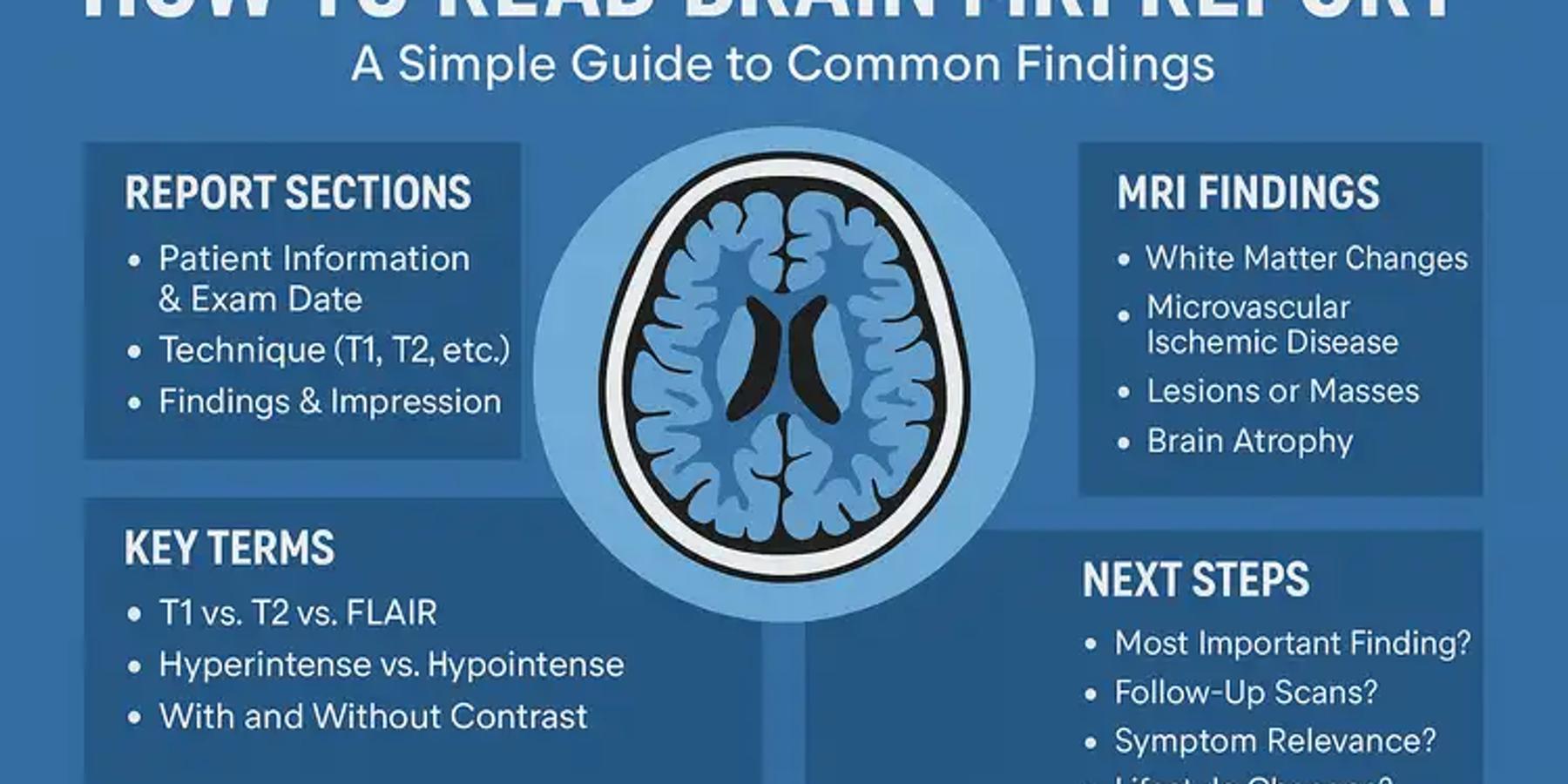
Almost every MRI report follows the same five-part structure, no matter which scan centre you go to or which city you are in. The five sections are Clinical History, Technique, Findings, Impression, and Clinical Correlation. Think of them as a story that moves from "why are we doing this scan" to "here is what we saw" to "here is the short version" to "go talk to your doctor."
Most patients panic in the Findings section, which is the longest and most technical, and skip the Impression, which is usually written in the clearest language. Reading them in order, knowing what each one is for, takes a lot of the fear out.
Clinical History
Clinical History is the short opening line that says why the scan was done. It usually repeats the symptom or the reason your doctor sent you, for example "headache for three months" or "knee pain after a fall." This is simply context: the radiologist noting the question they were asked to look into. It tells you nothing new about your results, so do not read meaning into it.
Technique
Technique is the section that describes how the scan was performed, which body part was scanned, and whether a contrast dye was used. Contrast is a special liquid, usually one called gadolinium, that is sometimes injected into a vein to make certain areas show up more clearly. If your report says "post contrast study," it simply means that dye was used. This section is mostly for the doctor's record. It confirms the right area was scanned in the right way. There is no hidden result here.
Findings
Findings is the long, detailed section where the radiologist describes everything they see, part by part. This is the section that scares people the most, because it lists every observation in technical language, including all the normal ones. A report will often spend several lines saying things look normal, written in words that do not feel reassuring at all, like "no focal lesion" or "normal signal intensity."
Read this section slowly. Remember that most of what is written here is the radiologist confirming that things are as expected. The few lines that describe something noticeable are not automatically bad news. They are observations your doctor will weigh. When you hit a word you do not recognise, do not spiral. The next part of this guide explains the common ones.
Impression
Impression is the summary section, and honestly it is the part you should read first. After describing everything in the Findings, the radiologist steps back and writes the short version: the few things that actually stood out and are worth your doctor's attention. It is usually only two or three lines, written more plainly than the rest. If you read nothing else carefully, read the Impression. Even here, though, it is a summary of observations, not a final diagnosis.
Clinical Correlation
Clinical Correlation is the closing line you will see on most reports, often phrased as "clinical correlation is advised" or "suggest clinical correlation." It sounds formal and slightly worrying, but it is one of the most reassuring lines in the whole report. It simply means the radiologist is saying their pictures need to be matched with your actual symptoms and examination before anything is concluded. In plain language: do not treat this report on its own, the treating doctor must connect it to you, the real person.
What do the common words in an MRI report mean?
Most of the fear in an MRI report comes from a small handful of repeating technical words. Almost all of them describe how something looks in the picture, not what is wrong with you. The words below describe brightness, scan settings, and appearance. None of them is a diagnosis on its own, and your doctor will decide what each means for you using your full clinical picture.
Signal, hyperintense, and hypointense
Signal is just the word for how bright or dark an area looks in the MRI picture. MRI images are shades of grey, and "signal" describes where on that grey scale something sits. Hyperintense means an area looks brighter or whiter than the tissue around it. Hypointense means an area looks darker than the tissue around it.
The simple memory trick: hyper is high and bright, hypo is low and dark.
On their own, neither is good or bad. Water and fluid often look bright, dense tissue often looks dark, and the radiologist uses these brightness differences to describe what they see. So "hyperintense signal" typically refers to an area that shows up brighter in the image. It is a description of appearance, not a diagnosis.
T1, T2, and FLAIR
T1, T2, and FLAIR are the names of different camera settings the MRI machine uses, not findings about your body. The same area gets photographed in a few different ways so the radiologist can compare them. The short version most patients find useful: on a T2 setting, fluid and water tend to look bright; on a T1 setting, fat tends to look bright. The radiologist looks at how an area behaves across both settings to understand what it might be made of.
FLAIR stands for Fluid Attenuated Inversion Recovery. That is a long name for a clever setting that dims down normal fluid so other things become easier to spot. So when your report mentions "T2 hyperintense" or "FLAIR hyperintense," it typically refers to how an area appears under a particular camera setting. These are descriptions of the image, not diagnoses, and your doctor reads them together with your symptoms.
Enhancement and contrast
Enhancement describes what happens after the contrast dye is given. If an area lights up or becomes brighter after the dye is injected, the report calls that enhancement. This happens because the dye travels in the blood, so areas with more blood supply tend to brighten. This is why "no abnormal enhancement" is usually a calm, reassuring phrase, even though it does not sound like one. It typically means nothing lit up in a way the radiologist found unusual.
Why should you not Google your MRI findings one word at a time?
There is a very Indian version of this trap, and it deserves naming. We do not sit quietly and wait days for the consultation. We share the report on the family WhatsApp group within minutes, where one uncle becomes a radiologist, one cousin becomes an oncologist, and someone forwards a frightening voice note from a person nobody has actually met. And because a second opinion is so easy to get here, we walk into a different doctor's clinic in the same neighbourhood within a couple of hours, often before we have understood the first report at all.
There is nothing wrong with a second opinion. But chasing one in a panic, armed with a half-understood word and a WhatsApp group's worst guesses, usually adds fear, not clarity. The healthier move is the opposite of word-by-word Googling: understand the whole report calmly, in plain language, then take your questions to a doctor who can connect the report to you.
What should you actually do after you read your MRI report?
After reading your report, the most useful thing you can do is slow down, read the Impression section first, and write down your real questions for your doctor instead of trying to diagnose yourself. The report is one input into a conversation, not the conversation itself. Your job is not to decode every line perfectly. Your job is to walk into your consultation understanding enough to ask good questions and actually hear the answers, instead of sitting there frozen by a word you read at midnight.
Here are calm, practical questions worth carrying to your doctor:
- In simple words, what does this report mean for me specifically?
- Which finding, if any, are we acting on, and which are we just noting?
- Does this change my treatment, or are we watching and waiting?
- Do I need any further tests, and if so, why?
- Is there anything reassuring in this report that I might have missed?
Bring the full report to the appointment, not a screenshot of one scary line. Give your doctor the whole picture, the same way you would want them to give you the whole picture. And if you genuinely cannot reach your doctor soon and the worry is unbearable, getting a plain-language explanation of the report first will at least let you wait without spiralling.
How FlexReport helps you understand your report
FlexReport is a patient comprehension layer for diagnostic centres, labs, and hospitals. It adds a plain-language second view to clinical reports without altering the clinical report itself. FlexReport operates in India and is built by FlexRad, a unified radiology platform with imaging infrastructure across India and the US.
In everyday terms: your radiologist's report stays exactly as written, because that is the official record and it should never be changed. FlexReport sits alongside it and adds a simplified explanation, in plain language, often on the same WhatsApp thread or patient app your diagnostic centre already uses. The terms get explained. The brightness words stop sounding like sentences. You get the calm, full-context version of your own report instead of the frightening fragment.
What FlexReport does not do is diagnose you, replace your doctor, or tell you what your treatment should be. That remains entirely with your treating physician, where it belongs.