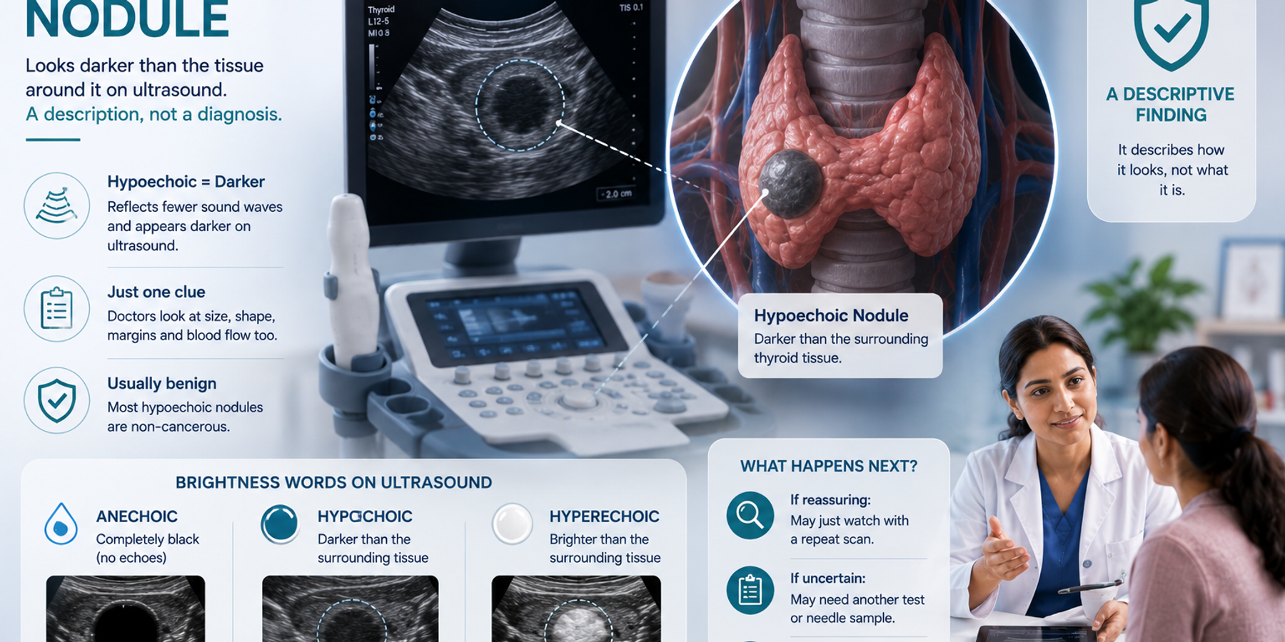
A hypoechoic nodule on your ultrasound simply means a lump that looks darker than the tissue around it on the scan, and the word describes only how it looks, not what it is. Let us break it down. A nodule is a small lump. Hypoechoic is made of hypo, meaning less or low, and echoic, meaning echoes. Ultrasound builds a picture from echoes, and an area that sends back fewer echoes appears darker on the screen. So hypoechoic just means darker than the surrounding tissue. It is one of several features radiologists note, alongside size, shape and edges, and on its own it does not tell you whether something is harmful. Most hypoechoic nodules are benign, meaning non-cancerous. On your report, hypoechoic is a description of what the scan showed, not a diagnosis. What it means for you is for your doctor to judge from the whole picture.
If the word hypoechoic on your report set your mind racing, slow down, because it is a plain description, not a warning. This guide explains what it means, how it fits with other features, and what to ask your doctor. And if the rest of the report reads like another language, FlexReport can help. Upload it on app.flexreport.ai and get a plain-language version in minutes.
What does hypoechoic mean?
Hypoechoic means an area looks darker than the tissue next to it on ultrasound because it reflects fewer sound waves back. Ultrasound turns echoes into shades of grey: tissues that bounce back more sound look brighter, and those that bounce back less look darker. Radiologists, the specialist doctors who read scans, use hypoechoic for these darker areas, hyperechoic for brighter ones, and anechoic for completely black, clear-fluid areas. None of these words is a diagnosis by itself. Hypoechoic is simply one clue among many, and radiologists always read it together with the nodule's size, shape, edges and blood flow before deciding whether it deserves a closer look (Radiological Society of North America).
Hypoechoic, hyperechoic, anechoic: the brightness words
Ultrasound reports lean on a small family of brightness words, and once you know them, they stop feeling like code. They describe only how light or dark something looks on the scan, which reflects what it is made of, not whether it is harmful. Clear fluid looks completely black, dense tissue looks bright, and many ordinary lumps look somewhere in between. The table translates the common ones so you can place whatever your report says.
| Word | How it looks | What it usually reflects |
|---|---|---|
| Anechoic | Completely black | Clear fluid, as in a simple cyst |
| Hypoechoic | Darker than nearby tissue | Denser or more solid than fluid, many benign causes |
| Isoechoic | Similar to nearby tissue | Blends in with normal tissue |
| Hyperechoic | Brighter than nearby tissue | Fat, calcium or dense tissue, often benign |
Hypoechoic just means darker on the scan. It is a clue, not a conclusion, and never a diagnosis on its own.
Is a hypoechoic nodule serious? Is it cancer?
A hypoechoic nodule is usually not serious and, on its own, does not mean cancer. Being darker on ultrasound is a common feature of many completely benign lumps. What actually guides your doctor is the whole set of features together: the nodule's size, shape, edges, whether it has blood flow inside, and whether it is growing. A hypoechoic nodule with otherwise reassuring features is often just watched, while one with several suspicious features may get a closer look. Even then, a closer look is not the same as a diagnosis. It means your doctor wants more information. A report line is one puzzle piece. Your doctor holds the whole puzzle.
What happens next after this finding?
What happens next depends on the full picture, not the word hypoechoic. If the nodule looks otherwise reassuring, your doctor may simply note it or suggest a repeat scan after some months to confirm it is stable. If several features together look less certain, the next step may be another test, such as a follow-up ultrasound, or a needle sample to check the cells, depending on the organ. The plan is guided by where the nodule is, since a hypoechoic thyroid nodule and a hypoechoic breast nodule are assessed with their own criteria. Follow-up scans are a normal way of keeping watch, not a sign that something is wrong.
A few useful questions to carry into your appointment:
- Apart from being hypoechoic, do the other features look reassuring?
- Do I need any further test, or can we watch it?
- When should I have a repeat scan?
FlexReport is a patient comprehension layer for diagnostic centers, labs, and hospitals. It adds a plain-language second view to clinical reports without altering the clinical report itself. FlexReport operates in India and is built by FlexRad, a unified radiology platform with DICOM and PACS infrastructure across India and the US.