Take a breath first, because an osseous abnormality is usually far less alarming than it sounds, and most are harmless. The word osseous simply means bony, or relating to bone, and abnormality just means something that looks different from the ordinary. So an osseous abnormality is nothing more than an area of bone that looks a little different from the normal bone around it on a scan. It is the same idea as a bone lesion or an osseous lesion, just worded differently. There are many everyday, non-cancerous reasons for one, from a harmless dense spot you were probably born with, to an old healed injury, to a simple fluid-filled cyst. Radiologists, the specialist doctors who read scans, use these neutral words to describe anything that stands out, not to raise alarm. On your report, an osseous abnormality is a description of what the scan showed, not a diagnosis. What it means for you is for your doctor to work out from the whole picture.
If a scan report just landed on your WhatsApp and the phrase osseous abnormality set your mind racing, slow down. This guide explains what the word means, why most are benign, how it relates to the term bone lesion, and what to ask your doctor. And if the rest of the report reads like another language, FlexReport can help. Upload it on app.flexreport.ai and get a simplified, plain-language version of the whole thing in minutes.
What does osseous abnormality mean?
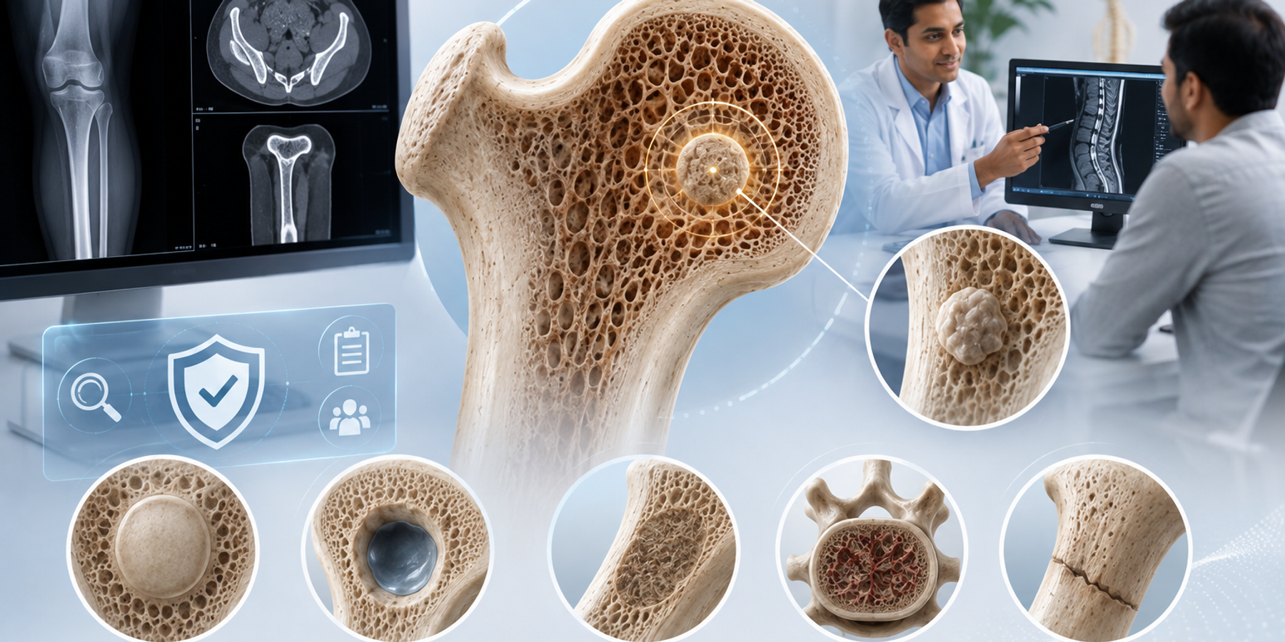
An osseous abnormality is an area of bone that looks different from the normal bone around it on an X-ray, CT or MRI scan. Osseous means bony, and the word does not say what caused the difference. It is a neutral placeholder that means something here looks a bit different, let us describe it. Bones are living tissue that constantly repair and remodel themselves, so they very commonly carry small marks: a dense spot called a bone island that many people are born with, the trace of an old injury that healed long ago, or a simple cyst, which is a harmless fluid-filled pocket. Radiologists note each of these because describing everything is their job, the way a surveyor lists every feature of a building. The presence of an osseous abnormality on your report is therefore not a verdict. It is the start of a description that your doctor and the radiologist read together (Radiological Society of North America).
Osseous abnormality, osseous lesion, bone lesion: the same idea
Reports use several words for the same basic idea, and they can be confusing when you first meet them, so it helps to see them side by side. Osseous abnormality, osseous lesion and bone lesion all describe an area of bone that looks different from normal. None of them carries a hidden, more serious meaning than the others. A report may also describe the appearance more precisely: a sclerotic area is one of denser, harder bone and is often a harmless bone island, while a lytic area is one where the bone looks thinner or emptier and has many benign causes but is reviewed on its features. Reading these as neutral descriptions, rather than as diagnoses, takes much of the fear out of a report. The table translates the common wordings. For a fuller walk-through of these findings, our companion guide on bone lesions goes into more detail.
| Words on your report | What they actually mean |
|---|---|
| Osseous abnormality | An area of bone that looks different from normal. Osseous means bony. |
| Osseous lesion / bone lesion | The same idea, worded differently. Most are harmless. |
| Sclerotic area | Denser, harder bone. Often a harmless bone island. |
| Lytic area | Bone that looks thinner or emptier. Many benign causes, reviewed on its features. |
| Well-defined / well-circumscribed | Clear, smooth edges, which is a typically reassuring feature. |
Osseous just means bony. An osseous abnormality is an area that looks different, and far more often it is ordinary than ominous.
Common benign causes of an osseous abnormality
Most osseous abnormalities picked up on everyday scans are benign, meaning non-cancerous, and radiologists recognise many of them on sight because they have such typical appearances. You do not need to memorise these, but seeing the common names in plain language can take the fear out of a report. A bone island is a small patch of extra-dense bone that is completely harmless. A simple bone cyst is a fluid-filled pocket. A non-ossifying fibroma is a very common harmless patch in children and teenagers that usually fades with age. A haemangioma is a harmless cluster of blood vessels, very common in the spine. An old healed fracture can also leave a mark that shows up as an abnormality long after it has mended. The table lists the frequent benign causes so you can match a name if your report gives one.
| Common benign cause | In plain terms |
|---|---|
| Bone island (enostosis) | A small patch of extra-dense bone. Harmless, often present for life. |
| Simple bone cyst | A fluid-filled pocket in the bone. Common, usually harmless. |
| Non-ossifying fibroma | A very common harmless patch in children and teens. Usually fades with age. |
| Haemangioma | A harmless cluster of blood vessels, very common in the spine. |
| Old healed fracture | The mark left by an injury that mended long ago. |
When does an osseous abnormality need a closer look?
An osseous abnormality gets a closer look based on its features and your symptoms, not on the word itself. Radiologists are reassured by areas with clear, smooth, well-defined edges that look the same on a repeat scan, because these are the hallmarks of the common benign causes. They pay more attention when an area has blurred or ragged edges, appears to involve nearby soft tissue, or grows or changes over time. Your own experience matters too: bone pain that is steadily worsening, that wakes you at night, or a bone that broke with very little force are all reasons for a prompt, careful review. The important thing to understand is that a closer look is not the same as a diagnosis. It simply means the radiologist wants more information, often just a comparison with an older scan, an MRI, or occasionally a biopsy where a tiny sample is examined. Most of the time, that closer look ends in reassurance.
One gentle note for the sons, daughters and spouses who often manage a family member's health, because in many Indian homes that is exactly who is reading this. Older people in particular tend to dismiss bone or joint pain as just age. If their report mentions an osseous abnormality, use it as a reason to ask, calmly, how they have really been feeling, and to book that visit. The word sounds scary, but the visit is usually reassuring.
What happens next after this finding?
What happens next depends on how the area looks and how you feel, and for most people it is low-key. When an osseous abnormality has all the classic features of a common benign cause, doctors very often do nothing at all, or simply compare it with older scans to confirm it has not changed. When the picture is less certain, the usual next step is more imaging, most often an MRI, which shows bone and soft tissue in more detail, or a comparison with a previous scan to check for change. In a smaller number of cases where the features are genuinely unclear, a doctor may recommend a biopsy, where a tiny sample is taken and examined so there is a definite answer rather than guesswork. A biopsy sounds daunting, but it exists to give certainty, and it very often brings reassuring news. Follow-up imaging is a normal way of keeping watch, not a sign that something bad has been found.
A few useful questions to carry into your appointment:
- Does my osseous abnormality look like one of the common benign causes?
- Do we have an older scan to compare it with?
- Do I need any further test, like an MRI, and if so, why?
- Which symptoms should bring me back to you sooner?
Walking in with your questions written on your phone keeps a five-minute consult from ending before your doubts do.
FlexReport is a patient comprehension layer for diagnostic centers, labs, and hospitals. It adds a plain-language second view to clinical reports without altering the clinical report itself. FlexReport operates in India and is built by FlexRad, a unified radiology platform with DICOM and PACS infrastructure across India and the US.