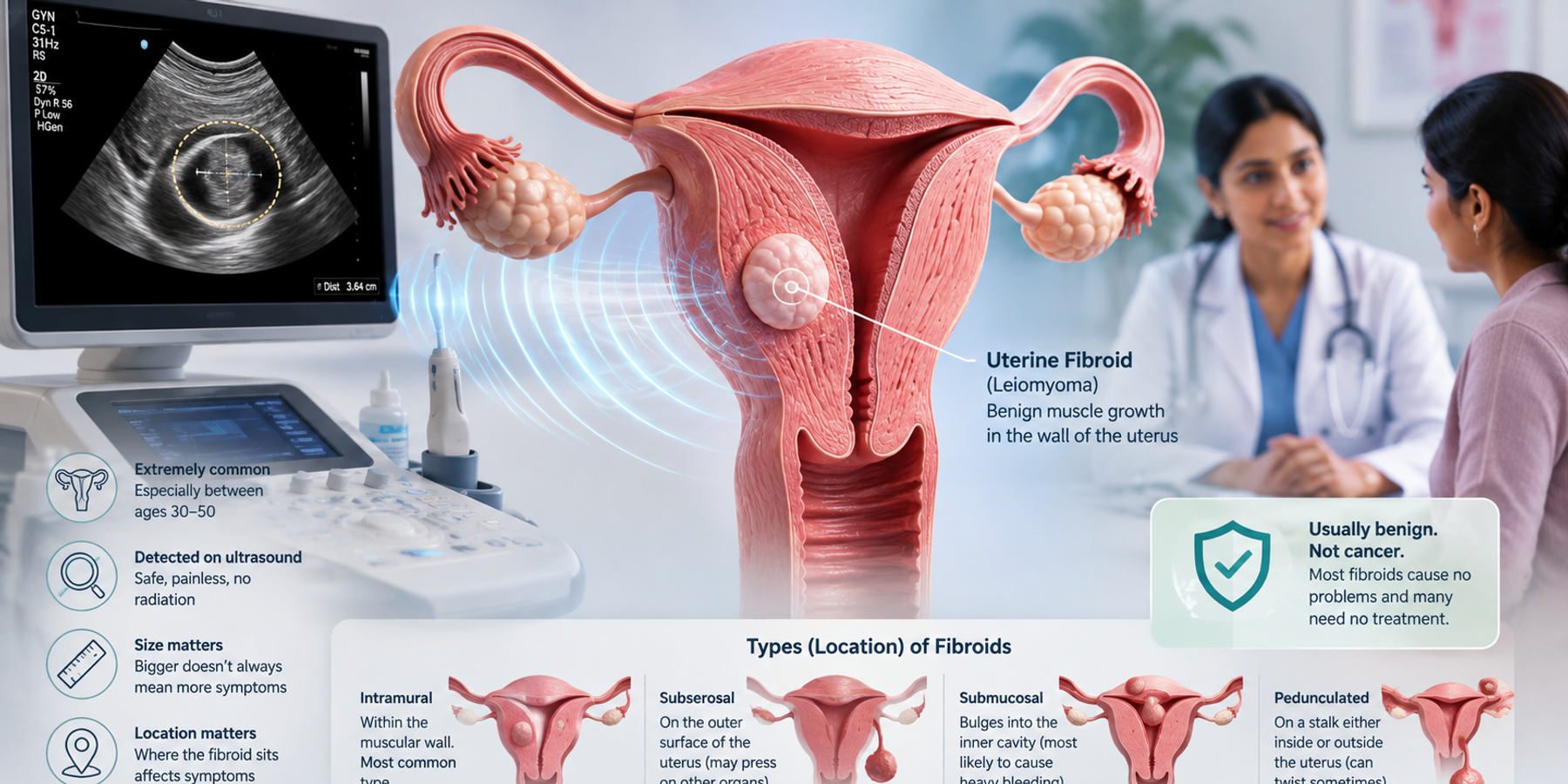
A uterine fibroid on your ultrasound usually means the scan found a harmless growth of muscle in the wall of your womb, and it is one of the most common findings in women. Let us break the words down. The uterus, or womb, is the organ where a baby grows during pregnancy. A fibroid is a firm lump made of the same muscle the uterus is built from, which doctors also call a leiomyoma or simply a myoma. An ultrasound uses sound waves, the same safe technology used to see a baby in pregnancy, to picture the uterus. So a uterine fibroid on ultrasound is a benign, meaning non-cancerous, muscle lump that the scan has found and measured. Fibroids are extremely common, especially between the ages of 30 and 50, and most cause no problems at all. What matters is not just the size but where the fibroid sits and whether it causes symptoms. On your report, the size and location are a description of what the scan showed, not a diagnosis.
If you are reading this because an ultrasound report just landed on your WhatsApp, maybe yours, maybe your wife's or your sister's, and a fibroid with a size in centimetres is worrying you, take a breath. This page explains what the size really tells you, what the different fibroid types mean, and what to ask your doctor. And if the rest of the report still reads like another language, FlexReport can help. Upload it on app.flexreport.ai and get a simplified, plain-language version of the whole thing in minutes.
What does a uterine fibroid on ultrasound actually mean?
A uterine fibroid on ultrasound is a benign lump of muscle in the uterus that the scan has detected and measured. Ultrasound is the main way fibroids are found because it is safe, painless and uses no radiation. Fibroids grow from the muscular wall of the uterus and are influenced by the body's natural hormones, which is why they are common during the reproductive years and often settle after menopause. A woman can have one fibroid or several, and they range from tiny seedlings to large lumps. Radiologists, the specialist doctors who read scans, describe each fibroid they see, including small ones that cause nothing, because their job is to record everything (Radiological Society of North America). The report will usually give the size in centimetres and often say where in the uterus the fibroid sits, since location affects symptoms as much as size does.
Here is the reassuring bit. A report noting a fibroid is extremely common and is not the same as your doctor being worried. Radiologists describe what they see, the way a home inspector notes every feature of a house, big or small. Fibroids are one of the most frequent notes they write. Listing one is duty. Deciding whether it needs anything is your doctor's job, and for a great many fibroids the answer is simply to keep an eye on it.
Uterine fibroid size on your report: a plain-English guide
The fibroid size on your report is simply how wide it measured on the scan, usually written in centimetres (cm) or millimetres (mm), where 1 cm equals 10 mm and one centimetre is about the width of a fingernail. Size is useful, but a bigger number does not automatically mean trouble, and a small fibroid in the wrong place can cause more symptoms than a large one sitting quietly. Doctors care about size mainly for two reasons: larger fibroids are more likely to cause pressure symptoms, and size helps guide which treatments make sense if any are needed. There is no single number that turns a harmless fibroid into a dangerous one, because fibroids are benign to begin with. The guide below shows how different sizes are commonly described, using everyday objects, but your doctor reads the size together with the location and your symptoms, which always comes first.
| Fibroid size | In everyday terms | How it is commonly viewed |
|---|---|---|
| Under 2 cm (under 20 mm) | Smaller than a grape | Small. Often just noted, frequently no symptoms. |
| 2 to 5 cm (20 to 50 mm) | Grape to a lime | Small to medium. Often watched. Symptoms depend on location. |
| 5 to 10 cm (50 to 100 mm) | Lime to an orange | Medium to large. More likely to cause symptoms, may be treated. |
| Over 10 cm (over 100 mm) | Larger than a grapefruit | Large. More likely to be discussed for treatment if it causes problems. |
Types of fibroids: why location matters as much as size
Fibroids are grouped by where they sit in the uterus, and this location often decides what symptoms, if any, they cause. Your report may use words like intramural, subserosal, submucosal or pedunculated, which sound technical but describe simple ideas. An intramural fibroid sits inside the muscular wall and is the most common type. A subserosal fibroid grows on the outer surface and can press on nearby organs such as the bladder, causing a need to pass urine often. A submucosal fibroid bulges into the inner cavity of the womb and, even when small, is the type most likely to cause heavy periods. A pedunculated fibroid hangs from a stalk, either inside or outside, and can occasionally twist and cause sudden pain. Knowing the type helps you understand your symptoms and helps your doctor choose the right plan. The table below translates the common types.
| Fibroid type | Where it sits | What it tends to mean |
|---|---|---|
| Intramural | Within the muscular wall | The most common type. May cause pressure or heavier periods if large. |
| Subserosal | On the outer surface | Can press on the bladder or bowel, causing frequent urination or fullness. |
| Submucosal | Bulging into the womb cavity | Most likely to cause heavy bleeding, even when small. Can affect fertility. |
| Pedunculated | On a stalk, inside or outside | Usually harmless, but can twist and cause sudden pain. |
With fibroids, where it sits can matter more than how big it is. A small one in the wrong spot outweighs a large one sitting quietly.
Is a uterine fibroid serious? Is it cancer?
The large majority of uterine fibroids are not serious and are not cancer. Fibroids are benign, meaning non-cancerous, growths, and cancerous change is very rare. Many fibroids cause no symptoms and are found by chance on a scan done for another reason. They matter when they cause heavy bleeding, pain or pressure, or affect fertility, and even then the goal is to manage symptoms, not to fear the fibroid itself. Doctors do pay closer attention to a fibroid that grows quickly, especially after menopause when fibroids normally shrink, because rapid change deserves a proper look, but this is uncommon. What matters is the full clinical picture: the size and location of the fibroid, your symptoms, your age and your plans for pregnancy. Assembling that picture is your doctor's job, not a search engine's. A report line is one puzzle piece. Your doctor holds the whole puzzle.
What symptoms can a fibroid cause?
Many fibroids cause no symptoms at all and are found only by chance. When they do cause symptoms, the common ones are heavier or longer menstrual periods, a feeling of pressure or fullness in the lower tummy, needing to pass urine more often, discomfort during sex, or lower back ache. Very heavy bleeding can sometimes lead to tiredness and low iron over time, which is worth mentioning to your doctor. None of these, on their own, mean anything dangerous, but they are worth treating because they affect daily life. There is a small set of warning signs that need prompt attention, because occasionally a fibroid can cause sudden severe pain or very heavy bleeding. The callout below spells out when to stop reading and get help.
One gentle note for the husbands, daughters and sisters who often help manage a family member's health, because in many Indian homes that is exactly who is reading this. Women frequently normalise heavy periods or pelvic pain and carry on, sometimes for years. If her report mentions a fibroid, use it as an opening to ask how heavy her periods really are and how she is feeling. It makes the doctor's visit far more useful.
What happens next after this finding?
What happens next depends on the fibroid's size and location, your symptoms and your stage of life, and for many women it is reassuringly simple. If a fibroid causes no symptoms, doctors very often just watch it, sometimes with a repeat scan after some months to check it is stable. If there are symptoms, the first steps are frequently medicines, such as treatments to reduce heavy bleeding or hormonal options that can ease symptoms or slow growth. When symptoms are significant, or a fibroid is large or affecting fertility, a gynaecologist may discuss procedures. These range from removing just the fibroid while keeping the uterus, called a myomectomy, to a uterine artery embolisation that shrinks fibroids by cutting their blood supply, up to a hysterectomy in more severe cases. There are many options between watching and surgery, so this is a conversation, not a sentence. Follow-up scans are a normal way of tracking a fibroid, not a sign that something bad has been found (Radiological Society of North America).
A few useful questions to carry into your appointment:
- What size is my fibroid, and where in the uterus does it sit?
- Do I need any treatment now, or can we watch it?
- Could this affect my periods or a future pregnancy?
- When should I have a follow-up scan, and which symptoms should bring me back sooner?
Walking in with your questions written on your phone keeps a five-minute consult from ending before your doubts do.
FlexReport is a patient comprehension layer for diagnostic centers, labs, and hospitals. It adds a plain-language second view to clinical reports without altering the clinical report itself. FlexReport operates in India and is built by FlexRad, a unified radiology platform with DICOM and PACS infrastructure across India and the US.