TLDR;
A fibroid is usually just watched when it is under 5 cm, taken more seriously once it crosses 5 cm, and treated as large once it is over 10 cm. But size is only half the story. Where the fibroid sits, and whether it is causing symptoms, matters just as much.
- Size is a starting point, not a verdict. Under 5 cm is often just monitored, 5 to 10 cm gets closer attention, and over 10 cm is usually treated.
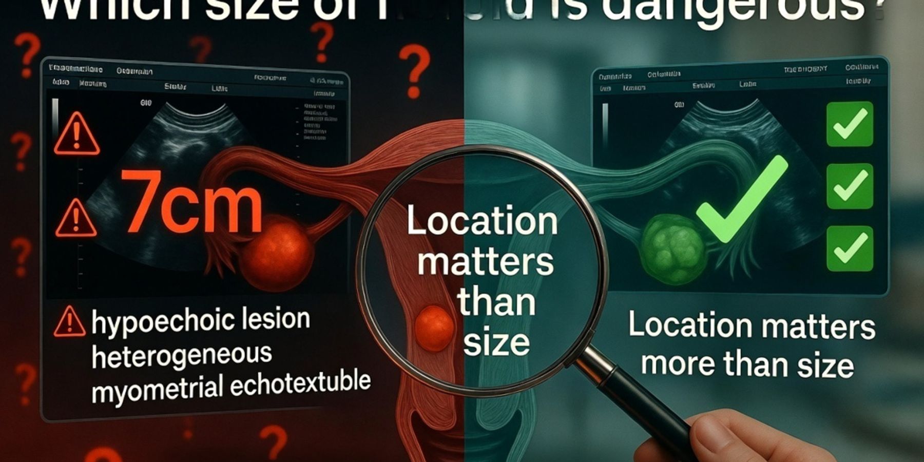
- Location can matter more than size. A small fibroid pressing into the inner lining of the uterus can cause more trouble than a much larger one sitting on the outer surface.
- Symptoms are what make a fibroid a problem, not the number. The ones doctors care about are heavy bleeding that leaves you tired and weak, pressure on the bladder or bowel, ongoing pain, fast growth, or trouble with pregnancy.
- Fibroids are almost always non-cancerous, and a large fibroid is not more cancerous than a small one.
- Best next step: take the actual report to a doctor in person. If your usual gynaecologist is busy, you can almost always see another good doctor in your area within a few hours.
This is a plain-language guide to help you understand your report. It is not a diagnosis. Your doctor decides what your specific fibroid means for you.
If you just got a scan report on WhatsApp and you are wondering which size of fibroid is dangerous, take a breath — the word next to that number is far less scary than it looks. This guide walks through it in plain language: what a fibroid actually is, how to read the size on your report (including the millimetre confusion that scares everyone), why location often beats size, when a fibroid genuinely becomes a problem, what the cancer question really deserves as an answer, and what to ask your doctor. No medical word is left unexplained.
This is a plain-language guide to help you understand your report — it is not a diagnosis, and your doctor decides what your specific fibroid means for you. And if at any point you want your own report turned into simple words, you can upload it to FlexReport for an easy-to-read second view. FlexReport does not change your report or replace your doctor; it just helps you understand what your scan says.
Which size of fibroid is dangerous? The quick answer
Doctors usually start watching a fibroid more closely once it grows past about 5 cm (roughly the size of a lemon), and they treat anything over 10 cm (about the size of a grapefruit) as large and more likely to need treatment. Below 5 cm, many fibroids cause no problems at all and are simply watched over time. That said, “dangerous” is not a line you cross at a fixed number. A fibroid becomes a real concern when it causes symptoms: heavy bleeding, pain, pressure on your bladder or bowel, or trouble with pregnancy. A 3 cm fibroid sitting inside the lining of your uterus can cause heavier bleeding than a 7 cm fibroid sitting quietly on the outer surface. So the size number on your report is a starting point, not a verdict — what your doctor really weighs is size, location, your symptoms, and how fast it is growing, all together.
First, what is a fibroid?
A fibroid is a non-cancerous growth made of muscle and fibrous tissue that develops in or on the wall of the uterus (the womb). You may also see it written on your report as a “leiomyoma” or “myoma,” which are simply the medical names for the same thing. The word “non-cancerous” is the important part: fibroids are not cancer, and they very rarely turn into cancer. They are also extremely common — by the age of 50, up to 80 percent of women will have developed fibroids at some point, and most never cause any symptoms at all.
Fibroids can be as small as a seed or grow as large as a melon, and a woman can have one fibroid or several at the same time. They tend to grow slowly, and many shrink on their own after menopause when hormone levels fall. None of this means you should ignore your report — it means the default situation is far less frightening than the word “tumour” makes it sound, because here a fibroid is a benign growth, not a dangerous one.
The fibroid size chart, in mm and cm
Your report almost certainly gives the size in millimetres (mm), while most articles talk in centimetres (cm), and the simple rule that fixes the confusion is this: 10 mm equals 1 cm. So if your report says a fibroid is 52 mm, that is 5.2 cm. If it says 18 mm, that is 1.8 cm — just move the decimal point one place to the left. Reports often give three numbers too, like “52 x 48 x 40 mm,” because a fibroid is a three-dimensional lump and the scan measures its length, width, and depth. Doctors usually focus on the largest of the three. Fibroids vary enormously, and can range from a few millimetres to more than 20 centimetres across.
To make the numbers feel real, here is a rough comparison many people find helpful:
| Size on report | In cm | Everyday comparison | General category |
|---|---|---|---|
| Up to 20 mm | Up to 2 cm | A blueberry or marble | Small |
| 20 to 50 mm | 2 to 5 cm | A grape to a lemon | Small to medium |
| 50 to 100 mm | 5 to 10 cm | A lemon to an orange | Medium |
| Over 100 mm | Over 10 cm | A grapefruit or bigger | Large |
These categories are a guide, not a strict rulebook. Different doctors draw the lines a little differently, and a number on its own never decides what happens next.
A quick size-by-size guide
Here is roughly what each common fibroid size tends to mean, keeping in mind that location and symptoms matter just as much as the number. This is a general guide to help you read your report calmly, not a diagnosis of your situation — only your doctor can tell you what your specific fibroid means for you.
| Fibroid size | Everyday comparison | What it usually means |
|---|---|---|
| Under 4 cm (under 40 mm) | A seed to a grape | Often causes no symptoms. Usually just watched over time. |
| 4 to 5 cm (40 to 50 mm) | A grape to a lemon | Many women have no symptoms. Usually monitored, treated only if it causes trouble. |
| 5 to 6 cm (50 to 60 mm) | Around a lemon | The rough point where doctors start watching more closely. May begin causing heavier periods or mild pressure. |
| 7 cm (about 70 mm) | A large lemon to a small orange | On the larger side. More likely to cause symptoms, and often discussed for treatment if it does. |
| 9 cm (about 90 mm) | Close to an orange | Large enough to press on nearby organs in some women. Frequently treated. |
| 10 cm and above (100 mm+) | A grapefruit or bigger | Usually treated, and more likely to need surgery, especially if it is causing symptoms. |
Why location matters more than size
Where a fibroid sits inside your uterus often matters more than how big it is — and this is the part almost no one explains. The uterus has three layers: an inner lining, a thick muscular middle wall, and a smooth outer surface. Where a fibroid grows decides what symptoms it causes, which is why two fibroids of the exact same size can behave completely differently. A small fibroid in a tricky spot can cause more trouble than a large one sitting out of the way. Your report may already name the location, so it helps to know what each type means. Doctors usually group fibroids by where they grow in the uterus.
The four types of fibroids, by location
A submucosal fibroid grows just under the inner lining of the uterus and bulges into the hollow space inside. These are the troublemakers — even when small, they tend to cause heavy or long periods, and they are the type most likely to affect fertility and pregnancy. An intramural fibroid grows inside the muscular wall itself; this is the most common type, and whether it causes problems depends mostly on its size and how many there are. A subserosal fibroid grows on the outer surface of the uterus and can balloon outward — these can grow quite large without causing heavy bleeding, but a big one can press on your bladder or bowel. A pedunculated fibroid grows on a thin stalk, a bit like a mushroom, either into the cavity or out from the surface; on rare occasions the stalk can twist and cause sudden pain, which is worth knowing but not worth losing sleep over.
So when you read your report, do not just look at the number — look for whether it says submucosal, intramural, subserosal, or pedunculated. A 2 cm submucosal fibroid may need more attention than a 6 cm subserosal one. This is a description of where the fibroid is, not a diagnosis of how serious your situation is; your doctor will use the location, alongside your symptoms and your full history, to decide what it means for you specifically.
How to read your own fibroid scan report
Your scan report is written for doctors, not for the person lying awake at 1 am trying to decode it — so here is what the most common words on it actually mean in plain language. Keep this section handy while you look at your own report. Everything below is the plain meaning of a term, not a judgement on your health.
If you see “bulky uterus,” it simply means your uterus is a little larger than the usual size, often because of fibroids or thickened muscle — a description, not an emergency. If you see “well-defined hypoechoic lesion,” break it down: “lesion” just means an area that looks different from the tissue around it (here, the fibroid), “well-defined” means it has a clear, neat border (which is reassuring), and “hypoechoic” is an ultrasound word meaning that area shows up darker on the scan, which is normal for fibroids. If you see “anteverted” or “retroverted,” that only describes the natural tilt of your uterus, forward or backward — a normal variation that has nothing to do with danger.
A fibroid is just one of many things that can show up on a scan. If your report mentions other words you do not recognise, our Radiology Findings Library explains them in the same plain language, finding by finding.
When a fibroid actually becomes a problem
A fibroid becomes a real problem when it causes symptoms, not when it reaches a particular size. The clearest warning sign is heavy periods that soak through pads or pass large clots, because steady blood loss month after month can lead to anemia, which is a low level of healthy red blood cells that leaves you tired, weak, and short of breath. Pressure symptoms are the next thing doctors watch, and large fibroids can press on the bladder and bowel, which can make you pass urine very often or cause constipation and a feeling of fullness. Ongoing pelvic pain, or pain during sex, also counts.
Two other situations get a doctor's quick attention. One is a fibroid that grows fast over a short period, because rapid growth is something they like to investigate even though it is usually still harmless. The other is a fibroid causing trouble with getting or staying pregnant. If you have any of these, that is a reason to see a doctor properly — not a reason to panic tonight. The symptoms are what matter, and symptoms can be managed.
Is my fibroid cancerous? The honest answer
Fibroids are almost always non-cancerous, and the size of your fibroid does not tell you whether it is cancer. A cancerous growth in the uterine muscle, called a leiomyosarcoma, is genuinely rare, and a regular fibroid turning into one is rarer still. A large fibroid is not “more cancerous” than a small one. The thing doctors actually keep an eye on is not size by itself but unusual, rapid growth — especially after menopause, when fibroids would normally be shrinking rather than growing.
So if your report shows a fibroid, even a big one, that on its own is not a cancer finding — it is a description of a common, benign growth. The responsible thing is to let your doctor track it over time rather than to assume the worst from a number on a phone screen. If you ever notice rapid growth, or new bleeding after menopause, mention it to your doctor promptly, because those are the situations worth checking properly.
Fibroids and pregnancy
Many women with fibroids have completely normal pregnancies, so a fibroid on your report does not automatically mean trouble ahead. Whether a fibroid affects pregnancy depends, once again, on its size and especially its location rather than the simple fact that it exists. Submucosal fibroids, the ones that bulge into the inner cavity of the uterus, are the type most likely to affect fertility or raise the chance of complications, because they take up the space where a pregnancy needs to settle. Fibroids sitting in the wall or on the outer surface often cause no problem at all.
A common worry is whether you can get pregnant with a 5 cm fibroid, and the reassuring reality is that many women do, particularly when the fibroid is not pushing into the cavity. If you are planning a pregnancy or are already pregnant, this is a good thing to raise with your gynaecologist so they can look at the specific location and decide whether anything needs attention. A fibroid and a healthy pregnancy are not opposites.
What happens next: watch, medicine, or surgery
For most fibroids, the first step is not surgery — it is simply keeping an eye on it. If a fibroid is small and not causing symptoms, doctors often choose “watchful waiting,” which means repeating a scan every few months to see whether it is growing or staying put. No treatment is needed for a quiet fibroid. When symptoms do appear, the next step is usually medicine aimed at managing them, for example tablets to reduce heavy bleeding, or hormone-based options that can ease symptoms and sometimes shrink the fibroid.
When a fibroid is large, growing, or causing symptoms that medicine cannot control, your doctor may discuss a procedure. The common ones are a uterine artery embolisation, which cuts off the blood supply so the fibroid shrinks; a myomectomy, which removes the fibroid while keeping your uterus intact (often the choice for women who want children); and a hysterectomy, which removes the uterus entirely and is usually a last resort. There is no single right answer for everyone, because the right treatment depends on your symptoms and the size and location of the fibroid. That is exactly why this is a conversation to have with your doctor rather than a decision to make from an article.
The Indian reality, and what to ask your doctor
In India you are rarely stuck waiting days for answers, so the best move is a calm, in-person visit with the actual report in hand. Here is how this usually plays out at home: the diagnostic centre sends your ultrasound report to your WhatsApp, you read words you have never seen before, and within minutes the report has been forwarded to three family groups where someone's neighbour's cousin had “the same thing.” By evening, a small fibroid has become a family crisis. The good news is that if your regular gynaecologist is not free, you can almost always see another good doctor in your area within a few hours. Use that.
A short conversation with a doctor will settle this faster than any family group ever will.
Before you go, write down a few questions so you make the most of the visit:
- How big is my fibroid, and where is it located? Is it submucosal, intramural, subserosal, or pedunculated?
- Is it causing my symptoms, or is something else going on?
- Does it need treatment now, or can we watch it?
- Will it affect my plans for pregnancy?
- What would make you want to see me again sooner?
Walking in with these questions turns a scary report into a clear plan. If you want to understand your report before that visit so your questions are sharper, you can get a plain-language version on FlexReport first.
FlexReport is a patient comprehension layer for diagnostic centres, labs, and hospitals. It adds a plain-language second view to clinical reports without altering the clinical report itself, on the same WhatsApp thread or app your centre already uses. FlexReport operates in India and is built by FlexRad, a unified radiology platform with DICOM/PACS infrastructure across India and the US.